Home
Uncategories
Compact Bone Diagram Lacunae - COMPACT BONE. if rings are less obvious look for the lacuna and bigger holes- haversian canals ... / Bone osseous tissue labeled cancellous bone structure spongy bone diagram compact bone connective tissue vascular lacunae compact bone 400x haversian system bone osteocyte function osteoblast osteocyte osteoclast bone cell.
Compact Bone Diagram Lacunae - COMPACT BONE. if rings are less obvious look for the lacuna and bigger holes- haversian canals ... / Bone osseous tissue labeled cancellous bone structure spongy bone diagram compact bone connective tissue vascular lacunae compact bone 400x haversian system bone osteocyte function osteoblast osteocyte osteoclast bone cell.
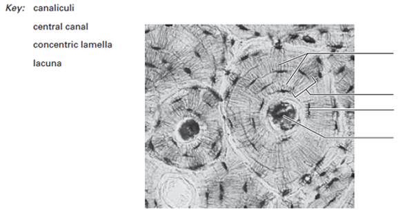
Compact Bone Diagram Lacunae - COMPACT BONE. if rings are less obvious look for the lacuna and bigger holes- haversian canals ... / Bone osseous tissue labeled cancellous bone structure spongy bone diagram compact bone connective tissue vascular lacunae compact bone 400x haversian system bone osteocyte function osteoblast osteocyte osteoclast bone cell.. It is a bone is one of two kinds of bone tissue that can be found in the body of a only tiny spaces (lacunae) are left which that contain the bone cells or osteocytes. Basically, in kindergarten when you drew skeletons, you were drawing compact bone. To know the architecture of compact and spongy (cancellous) bone. Learn vocabulary, terms and more with flashcards, games and other study tools. Lacunae, small chambers containing osteocytes, are arranged concentrically around the central canal.
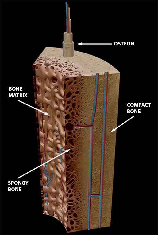
Compact bone forms the surface of all bones. Edraw is a new uml diagram and software diagram drawing tool. Like compact bone, spongy bone, also known as cancellous bone, contains osteocytes housed in lacunae, but they are not arranged in concentric circles. To know the architecture of compact and spongy (cancellous) bone. Find stockbilleder af anatomy compact bone i hd og millionvis af andre royaltyfri stockbilleder, illustrationer og vektorer i shutterstocks samling.
Lacuna Anatomy - Anatomy Drawing Diagram from media.cheggcdn.com Osteocytes occupy spaces (lacunae) in the bone matrix. The walls of the diaphysis are composed of dense and hard compact bone. Thin layer of reticular ct lining internal marrow cavity. Osteoblasts deposit the matrix in the form of thin sheets which are called lamellae. Compact bone is dense bone tissue found on the outside of a bone. In an ordinary microscopic section, viewed by transmitted light, they appear as fusiform opaque spots. Once the osteoid is mineralized, the precursor cells get surrounded by organic intracellular substances called lacunae to become fully developed and matured into osteocytes. Compact bone also called cortical bone dense bone in which the bony matrix is solidly filled with organic ground substance and inorganic salts leaving only tiny spaces lacunae that contain the osteocytes or bone cells.
Bone osseous tissue labeled cancellous bone structure spongy bone diagram compact bone connective tissue vascular lacunae compact bone 400x haversian system bone osteocyte function osteoblast osteocyte osteoclast bone cell.
Tusindvis af nye billeder af høj kvalitet tilføjes hver dag. Start studying compact bone structure. It is a bone is one of two kinds of bone tissue that can be found in the body of a only tiny spaces (lacunae) are left which that contain the bone cells or osteocytes. Blood vessels and nerves enter the bone through. Spongy and compact bone diagram / bone … long bone diagram trabeculae : A bone is a rigid tissue that constitutes part of the vertebrate skeleton in animals. The walls of the diaphysis are composed of dense and hard compact bone. Compact bone diagram bone cross section diagram file624 diagram of compact bone new. Learn vocabulary, terms and more with flashcards, games and other study tools. To know the structures of a synovial joint and a symphysis joint (intervertebral disc). Compact bone also called cortical bone dense bone in which the bony matrix is solidly filled with organic ground substance and inorganic salts leaving only tiny spaces lacunae that contain the osteocytes or bone cells. Bone diagram labeled data wiring diagram today. Compact bone consists of closely packed osteons or haversian systems.
Compact bone tissue diagram quizlet. Bone osseous tissue labeled cancellous bone structure spongy bone diagram compact bone connective tissue vascular lacunae compact bone 400x haversian system bone osteocyte function osteoblast osteocyte osteoclast bone cell. The basic units of compact bone are called osteons or haversian systems. Lesion composed of dense cortical bone (compact bone) with definite osteocyte lacunae and cement lines (line visible by microscopic examination marking the boundary of an osteon/ haversian system). In histology, a lacuna is a small space, containing an osteocyte in bone, or chondrocyte in cartilage.
Compact Bone Diagram - koibana.info | Cell diagram, Anatomy and physiology, Anatomy bones from i.pinimg.com A bone is a rigid tissue that constitutes part of the vertebrate skeleton in animals. Blood vessels and nerves enter the bone through. It can be remodeled all throughout life to withstand stress. Lacunae, small chambers containing osteocytes, are arranged concentrically around the central canal. Diagram of blood and nerve supply to bone. Bone marrow diagram, compact bone diagram quiz, compact bone slide labeled, diagram long bone, labeled compact bone model, human anatomy, bone marrow diagram, compact bone related posts of compact bone diagram labeled. Like compact bone, spongy bone, also known as cancellous bone, contains osteocytes housed in lacunae, but they are not arranged in concentric circles. Compact bone is dense bone tissue found on the outside of a bone.
Bone canaliculi are microscopic canals between the lacunae of ossified bone.
A bone is a rigid tissue that constitutes part of the vertebrate skeleton in animals. The walls of the diaphysis are composed of dense and hard compact bone. Canaliculi allow the passage of interstitial fluid between the central canal and the lacunae housing osteocytes. Compact bone diagram osteon compact bone ap pinterest anatomy human anatomy and. Compact bone is dense bone tissue found on the outside of a bone. Compact bone forms the surface of all bones. Compact bone tissue diagram quizlet. Compact bone surrounds the spongy bone tissue and it has a unique appearance. Osteoblasts deposit the matrix in the form of thin sheets which are called lamellae. Compact bone diagram osteon compact bone ap pinterest anatomy human anatomy and. You should include the histology of compact bone slides with diagram as well into your article. Compact bone also called cortical bone dense bone in which the bony matrix is solidly filled with organic ground substance and inorganic salts leaving only tiny spaces lacunae that contain the osteocytes or bone cells. A structural unit of compact bone consisting central haversian canal.
These cells are responsible for. The osteocytes are sitting in the lacunae and the canals are canaliculi, which interconnect the lacunae with the major vessels. In an ordinary microscopic section, viewed by transmitted light, they appear as fusiform opaque spots. Thin layer of reticular ct lining internal marrow cavity. Diagram of blood and nerve supply to bone.
In The Diagram Where Is The Osteon - Atkinsjewelry from www.visiblebody.com Compact bone consists almost entirely of extracellular substance, the matrix. Osteoblasts deposit the matrix in the form of thin sheets which are called lamellae. Compact bone forms the surface of all bones. Thin layer of reticular ct lining internal marrow cavity. Bone canaliculi are microscopic canals between the lacunae of ossified bone. It can be remodeled all throughout life to withstand stress. Each haversian canal is surrounded by concentric rings of compact bone called lamellae. Compact bone tissue diagram quizlet.
Compact bone is sometimes called cortical bone.
Compact bone diagram osteon compact bone ap pinterest anatomy human anatomy and. The osteocytes are sitting in the lacunae and the canals are canaliculi, which interconnect the lacunae with the major vessels. Tusindvis af nye billeder af høj kvalitet tilføjes hver dag. These cells are responsible for. You should include the histology of compact bone slides with diagram as well into your article. Like compact bone, spongy bone, also known as cancellous bone, contains osteocytes housed in lacunae, but they are not arranged in concentric circles. Compact bone consists almost entirely of extracellular substance, the matrix. The basic units of compact bone are called osteons or haversian systems. Bone canaliculi are microscopic canals between the lacunae of ossified bone. A diagram of a section of compact bone showing haversian canals. 6 compact bone vs spongy bone. The lacunae are situated between the lamellae, and consist of a number of oblong spaces. To know the architecture of compact and spongy (cancellous) bone.
Once the osteoid is mineralized, the precursor cells get surrounded by organic intracellular substances called lacunae to become fully developed and matured into osteocytes compact bone diagram. Compact bone is dense bone tissue found on the outside of a bone.

0 Comments:
Posting Komentar